

How is Plantar Fasciitis Diagnosed?

Sharp heel pain in the morning; tenderness at the bottom of the heel; stiff heels - these can all be signs of plantar fasciitis. However, without a proper diagnosis from a professional, it will just be guesswork.
In this article, we’ll explain how plantar fasciitis is actually diagnosed and whether imaging such as X-ray and MRI is required.
An Overview
Plantar fasciitis is diagnosed primarily through a patient’s history, symptoms and a physical examination.
Here’s how healthcare providers typically make the diagnosis:
1. Medical History
Your clinician/podiatrist will ask questions such as:
- Where exactly is the pain? (Typically at the bottom of heel, near the arch)
- When does it hurt most? (Often worst with first steps in the morning or after rest)
- What activities worsen it? (Impact and prolonged pressure activities such as standing, walking and running)
- Have you recently increased activity or changed footwear?
These patterns alone are often very characteristic of plantar fasciitis.
2. Physical Examination
A provider will usually check for:
- Localised heel tenderness – Pain is usually sharp when pressing directly over the medial calcaneal tuberosity (the large, prominent posterior part of the heel bone) which is the origin of the plantar fascia.
- Windlass Test – The toes are dorsiflexed (pulled upward). If pulling the toes upward causes heel and/or arch pain, it's considered a positive sign.
- Tight calf muscles – Limited ankle dorsiflexion (tight Achilles tendon) is common and increases strain on the plantar fascia.
- Gait assessment – Foot conditions such as flat feet, high arches, overpronation or any altered walking patterns may indicate plantar fasciitis.
3. Imaging (Usually Not Needed)
Imaging is only used if:
- Symptoms do not improve after several months
- Diagnosis is uncertain
- Concern for other conditions (e.g. stress fracture, nerve entrapment, tear etc.)
The most common types of imaging techniques used are: - Ultrasound – Most sensitive for plantar fasciitis and shows thickened plantar fascia (>4 mm).
- X-ray – Typically normal; may show a heel spur which does not cause the pain but sometimes appears in long-term cases.
- MRI – Rarely needed and only used if ruling out a fascia tear or complex heel pain.
Similar Conditions
There are other foot conditions that can mimic plantar fasciitis, and many of them will exhibit the same symptoms.
Therefore, a clinician might rule out:
- Heel stress fracture
- Tarsal tunnel syndrome
- Fat pad atrophy
- Achilles tendinopathy
- Systemic inflammatory conditions
Our Best Plantar Fasciitis Insoles
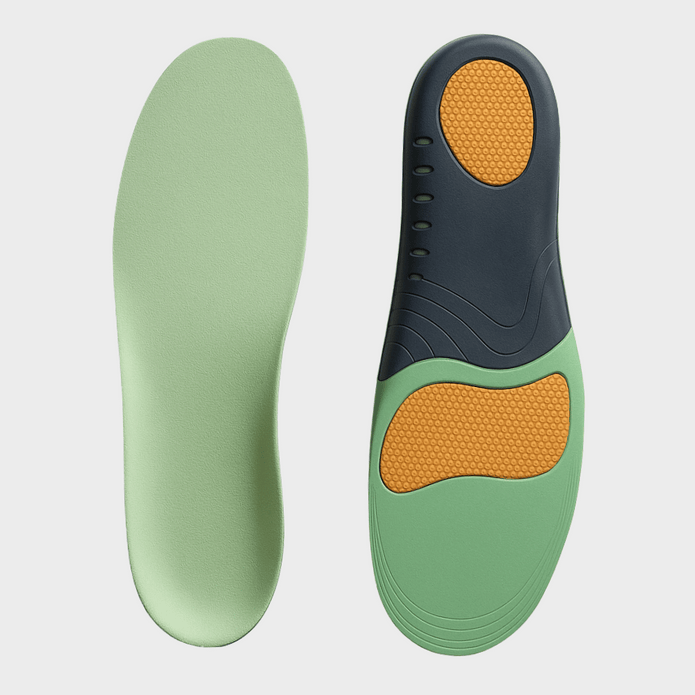

UltraSupport Plantar Fasciitis Insoles
View Product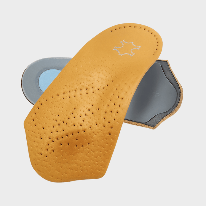
UltraSupport Plantar Fasciitis Leather Insoles
View ProductWhat Happens After?
Once plantar fasciitis is diagnosed, the next steps focus on reducing pain, promoting healing and preventing recurrence. Most people improve with conservative (non-surgical) treatment, especially when started early.
Here’s what typically what’s recommend by professionals after diagnosis:
- Education & Activity Modification – You’ll usually be advised to reduce high-impact activities temporarily and switch to low-impact exercise (cycling, swimming), among other effective recovery measures. Understanding what triggers your pain is a major part of recovery.
- Stretching & Flexibility Program – This is the cornerstone of treatment and aims to help regain strength and mobility.
- Footwear & Orthotics – Your provider may recommend supportive shoes with cushioned heels, over-the-counter insoles, heel cups or gel inserts for shock absorption. Custom orthotics is sometimes recommended if OTC supports aren’t enough.
- Pain Management – Methods to manage pain usually includes NSAIDs (ibuprofen, naproxen) to reduce pain and inflammation and/or ice therapy.
- Physical Therapy – If symptoms persist, physical therapy can add manual stretching and soft tissue techniques, strengthening of intrinsic foot muscles, gait correction, taping techniques and eccentric calf strengthening.
- Night Splints (If Needed) – These will keep the ankle slightly flexed overnight to prevent the fascia from tightening while you sleep and reduce severe “first-step pain” in the morning. This is recommended especially for chronic cases.
- Advanced Treatments – If conservative treatments fail (e.g. if pain lasts more than 6-12 months), providers may consider corticosteroid injections, shockwave therapy (ESWT), Platelet-rich plasma (PRP) injections or surgery (very rare and only considered when all other treatments fail).
- Monitoring Progress – Most cases improve over 6–12 weeks, with full recovery in 3–12 months, depending on severity and consistency with care. Your provider may check your gait mechanics, reassess footwear and adjust your treatment plan based on how you respond to treatments.
If you’re experiencing any symptoms that are typically associated with plantar fasciitis, it’s always advised to see a clinician/podiatrist to get a proper diagnosis rather than making assumptions.


Leave a comment